678
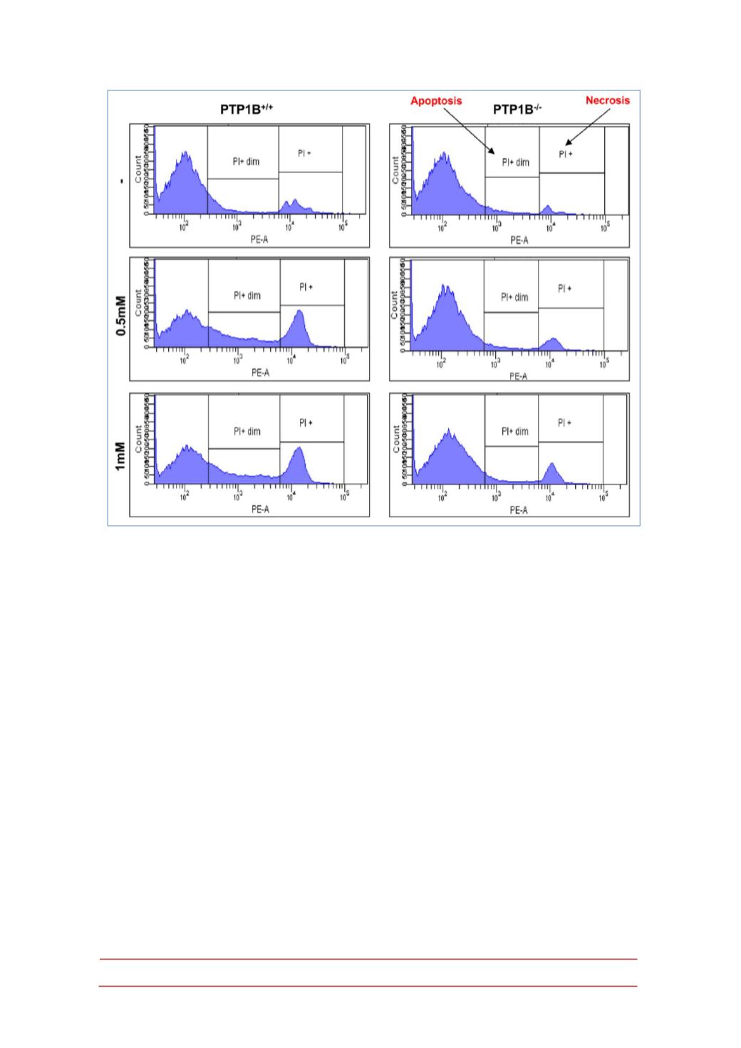
Figure 6.- PTP1B-deficient hepatocytes are protected against APAP-induced apoptosis and
necrosis.
PTP1B
+/+
and PTP1B
-/-
immortalized
hepatocytes were treated with various doses of
APAP for 16 h. Apoptosis and necrosis was measured by PI staining by flow cytometry (n=4
independent experiments).
To study the effect of APAP on the survival signaling pathways,
phosphorylation of the IGF-IR, levels of IRS1 and activation of Akt were examined.
As depicted in Figure 7B, phosphorylation of the IGF-IR and its downstream target
Akt decreased in APAP-treated wild-type hepatocytes at 8 h, but it was maintained
along APAP treatment in PTP1B-deficient hepatocytes. These results indicate that
the protective effect of PTP1B deficiency also involve the increase in survival
signaling. Interestingly, in PTP1B-deficient hepatocytes IRS1 degradation induced
by APAP was attenuated. Alltogether these results suggest that the increased
tyrosine phosphorylation of IGF-IR as a result of PTP1B deficiency elicits
hepatoprotection in conjunction with the attenuation of stress-mediated signaling.
Bcl-xL is a member of the Bcl-2 family with anti-apoptotic properties (40-
41). On that basis, we analyzed the expression of Bcl-xL after 8 h of APAP
treatment in wild-type and PTP1B
-/-
hepatocytes. As shown in Figure 7C, wild-type
cells showed a decrease in the expression of BclxL after APAP treatment whereas
BCLxL expression was maintained in hepatocytes lacking PTP1B.


