Protein tyrosine phosphatase deficiency…
673
susceptibility of hepatocytes to undergo apoptotic cell death induced by APAP. As
indicated in the introduction section, APAP overdoses cause severe hepatotoxicity
leading to liver failure in experimental animals and humans.
APAP hepatotoxicity
is, in part, the result of a series of events that increase cellular oxidative stress. In
the liver, Cyp2e1 converts APAP to NAPQI that rapidly depletes GSH and the
subsequent generation of ROS (7), and therefore the degree of GSH consumption is
a biomarker for APAP bioactivation (35). Both the catalytic and the modifier
subunits of γ-glutamyl cysteine ligase (GCL-C and GCL-M) are responsible for
glutathione synthesis. Since the expression of Cyp2e1, GCL-C and GCL-M did not
change in primary and immortalized hepatocytes from both genotypes of mice
(Figure 2A), we used immortalized cells for further experiments.
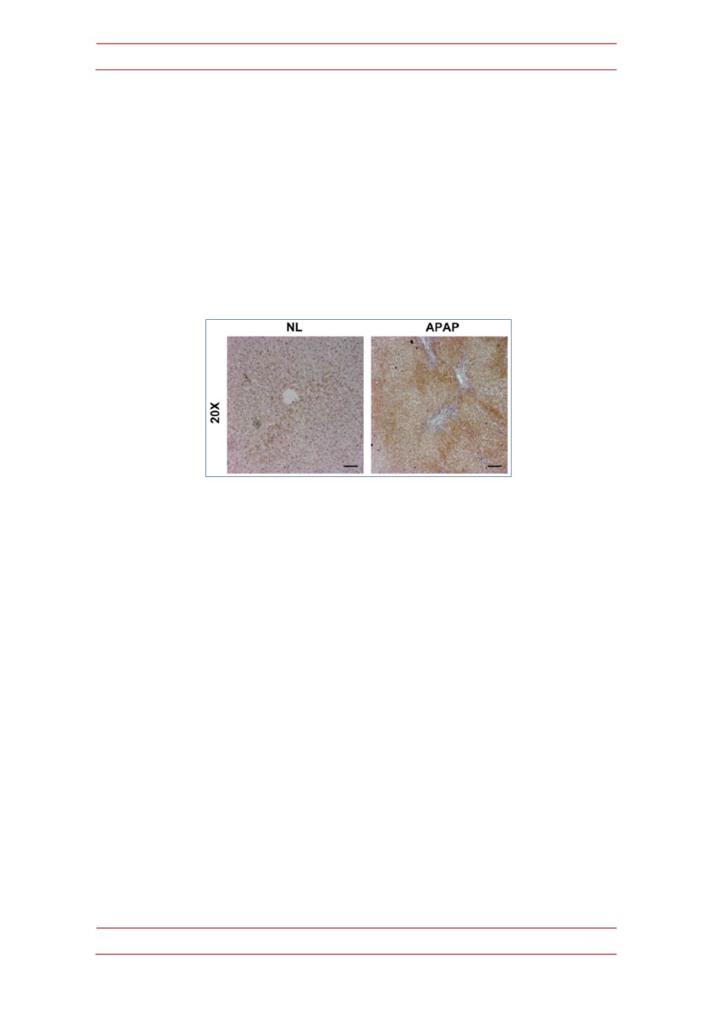
Figure 1.-PTP1B expression is increased during APAP-induced liver injury.
Representative
anti-PTP1B immunostaining of liver biopsy sections from an individuals with histologically normal
liver (NL) or with APAP overdose intoxication (APAP). Bar scale 50 µm.
It has been extensively reported that APAP hepatotoxicity concurs with
elevated ROS (36). To evaluate the degree of cellular oxidative stress in APAP-
treated hepatocytes, the intracellular ROS production was estimated. Figure 2B
shows a representative plot with the shift of the mean fluorescence after APAP
treatment for 6 h at 0.5 and 1 mM concentrations in immortalized hepatocytes.
Importantly, ROS production was significantly ameliorated in hepatocytes lacking
PTP1B compared to the wild-type controls.
Programmed cell death (apoptosis) induced by APAP treatment involves
activation of caspase-3: protective effect of PTP1B deficiency
.
To further investigate whether caspase-3 is involved in APAP-induced
apoptosis, we examined caspase-3 activity in wild-type and PTP1B
-/-
immortalized
hepatocytes. Cells were treated with APAP in a dose-dependent manner and
caspase-3 enzymatic activity was analyzed as described in Materials and Methods.
As shown in Figure 3A, caspase-3 activity increased with APAP treatment for 8 h in
wild-type hepatocytes with a maximal effect at 1 mM concentration. However, in
the absence of PTP1B, hepatocytes were protected against activation of caspase-3
upon APAP treatment.


