676
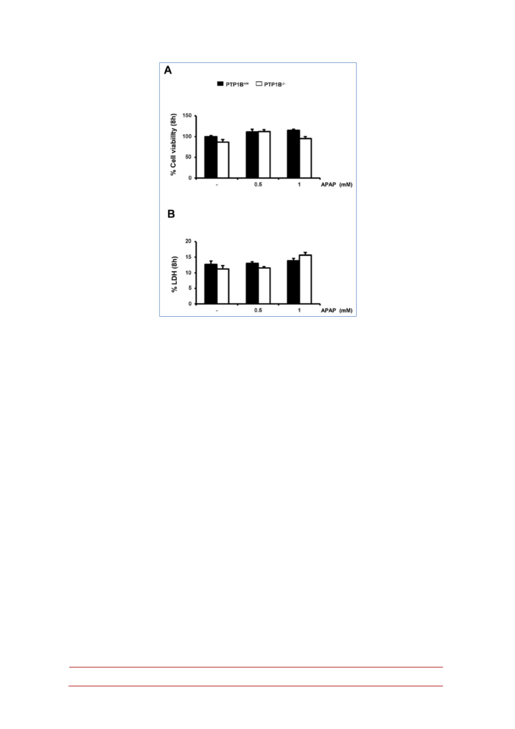
Figure 4.- Immortalized PTP1B-deficient hepatocytes are protected against APAP-induced
cell death.
PTP1B
+/+
and PTP1B
-/-
immortalized hepatocytes were treated with various doses of
APAP for 8 h. Cellular viability (A) and released LDH activity (B) were analyzed.
*
P<0.05,
**
P<0.01
and
***
P<0.005 PTP1B
-/-
vs. PTP1B
+/+
hepatocytes (n=4 independent experiments).
Cell death (irreversible loss of vital cellular structure and function) is a
fundamental phenomenon of biological organisms. Several lines of investigation
have led to the concept that there are two fundamental types of cell death:
apoptosis and necrosis. The findings of our study demonstrated that the
administration of APAP at heptotoxic doses led to the induction of cell death by
both necrosis and apoptosis, with apoptotic cell death typically preceeding
necrosis. Therefore, we analyzed the population of apoptotic and necrotic cells in
response to APAP by flow cytometry. The results shown in Figure 6 and Table 1
indicate a protection against both types of cell death in response to APAP in
PTP1B
-/-
hepatocytes as compared to wild-type cells.
Effect of APAP treatment in the signaling pathways that modulate cell death
and survival in wild-type and PTP1B-deficient hepatocytes.
At the molecular level, we analyzed both stress-mediated and survival
signaling in wild-type and PTP1B
-/-
hepatocytes upon APAP treatment. The c-Jun
N-terminal Kinases (JNKs), a subfamily of the mitogen-activated protein (MAP)
kinases, have been shown to be activated by phosphorylation at early time-periods
after APAP treatment in hepatocytes (38-39). To analyze JNK activation in
immortalized hepatocytes, cells were treated with different doses of APAP for 8 h
and JNK phosphorylation was examined by Western blot analysis. As shown in


